Dental Implants in Türkiye
Titanium roots that restore a single tooth or a whole jaw, placed with 3D-guided planning.
Learn more
Patient results
Every photograph on this page is a patient treated at this clinic in Ankara. Nothing here is stock photography and nothing has been retouched.
Before-and-after photography is the easiest thing in dentistry to make dishonest, so it is worth saying plainly how these were taken and what they do and do not tell you.
They are our own patients. Every image was taken at the clinic, not bought from a library and not borrowed from a supplier's catalogue. Each one is published with the patient's written consent.
They are unedited. No filters, no whitening in software, no smoothing of gums or edges. What the camera recorded is what you see.
Where the two shots do not match, we say so. Some pairs were taken the same way twice, with the same cheek retractors and the same lighting; those are the ones you can compare most directly. Others pair a retracted clinical view with a natural smile, because that is what the patient came back with. Each case tells you which it is, because a before shot in harsh clinical framing next to a relaxed, well-lit smile flatters the result on its own.
What they cannot tell you is what you would get. Bone, gums, bite and starting shade differ from mouth to mouth, and none of them can be assessed from a photograph of somebody else. If a case here resembles your situation, send us a photograph of your own teeth and we will tell you what is realistic before you book anything.
 Before
 After
After
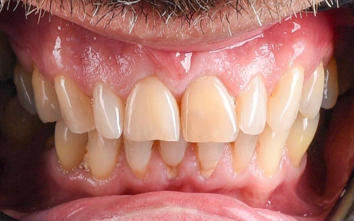
Years of wear had shortened the upper front teeth until their edges no longer followed the curve of the lower lip. The shade had darkened unevenly, old restorations no longer sealed at the margins, and the gum was visibly inflamed where they met the tooth. In the lower arch the front teeth were crowded and worn flat at the tips.
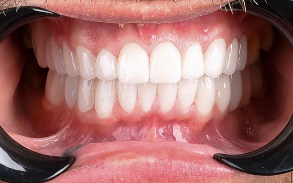
Both arches were rebuilt in ceramic. Length and proportion were returned to the upper front teeth so the incisal line follows the lip again, the shade was taken to a single consistent value across both arches, and the margins were finished so the gum could settle cleanly against them.
 Before
 After
After
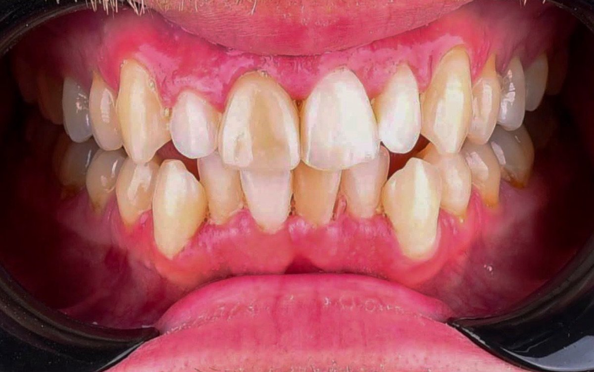
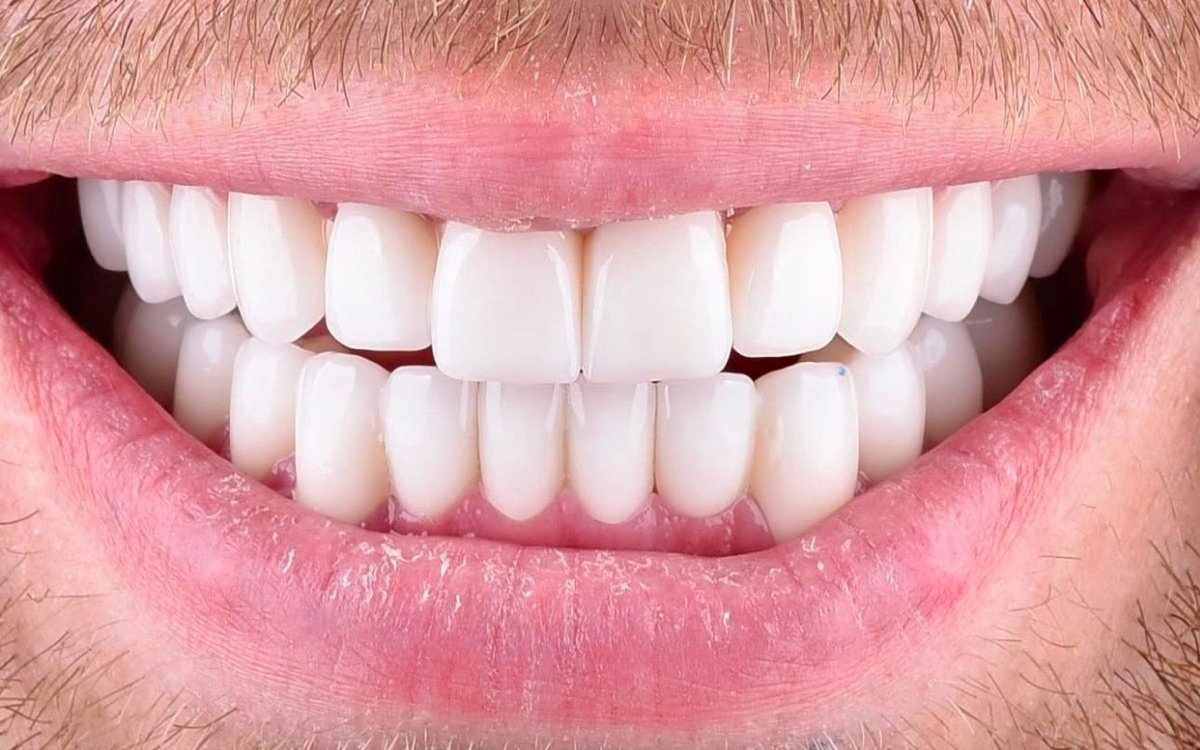
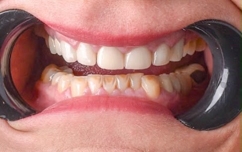
Before treatment the upper arch did not read as one colour: the two central incisors were noticeably lighter than the teeth beside them, which had darkened towards yellow-brown. The lateral incisors sat rotated and set back behind the centrals, the incisal edges stepped up and down instead of following a line, and the gum margin above the front teeth was red and swollen. The lower front teeth were crowded and overlapping, with a darker shade and an uneven edge line of their own.
After treatment both arches read as a single shade from one side to the other, the upper front teeth follow one incisal line instead of stepping, and the teeth sit along the arch rather than rotated behind one another. The after photograph is a natural smile rather than a retracted view, so the two images are not framed the same way: the gum line you can see in the first photograph is largely covered by the lip in the second.
 Before
 After
After
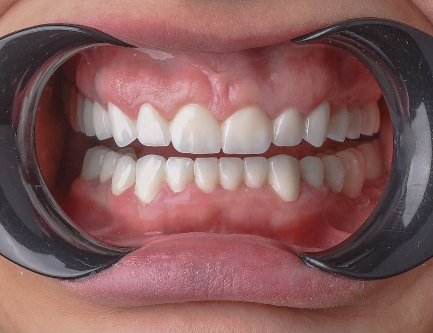
The lower front teeth had discoloured to a deep brown along the gum line, and their edges had worn down into an irregular line. The upper teeth were paler but no longer matched them, so the two arches read as two different colours whenever the mouth opened.
Both arches were brought to a single shade and the edges back to an even line. In the second photograph the gum margins sit cleanly against the restorations. Both images are retracted views, so what changed can be compared directly.
 Before
 After
After
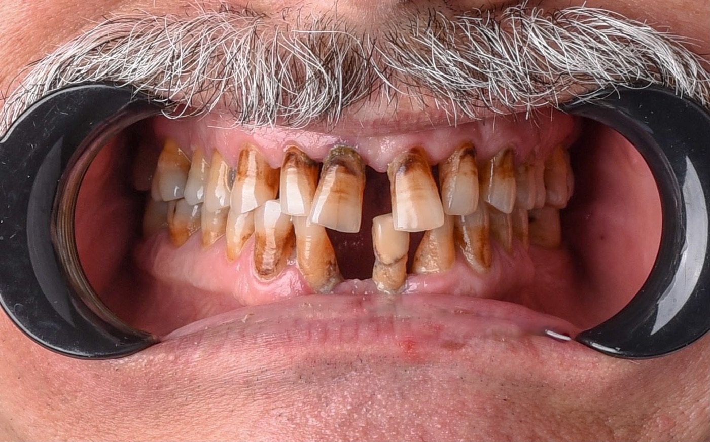
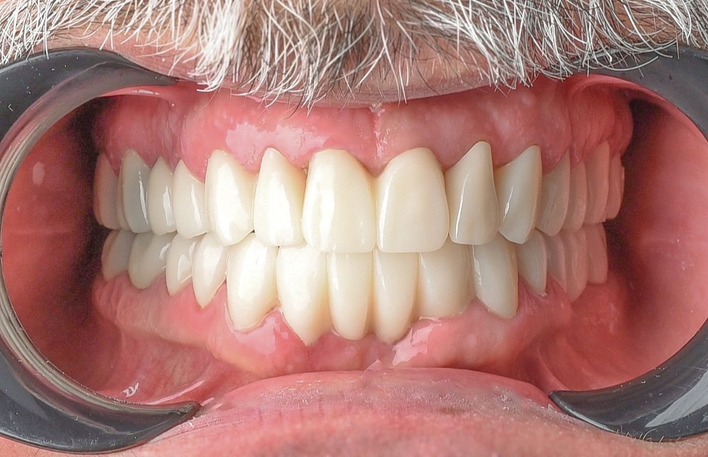
This is the most advanced case on the page. Nearly every visible surface was affected: the teeth were stained dark brown, worn short and irregular, gaps had opened between the front teeth, and the gum had receded far enough to expose root surface on several of them.
Both arches were rebuilt. The photograph after treatment shows an even edge line, one consistent shade across both arches, and gum margins settled against the restorations. Both images are retracted views taken the same way.
The part worth knowing is what was not done. A mouth in this state looks like a candidate for clearing and starting again, and it would have been quoted that way in plenty of places. In the event, one tooth was extracted and one tooth needed root canal treatment. Everything else was kept. Natural roots hold bone in a way that nothing replacing them does, and a tooth that can be saved is worth more than the tidiest possible plan.
 Before
 After
After
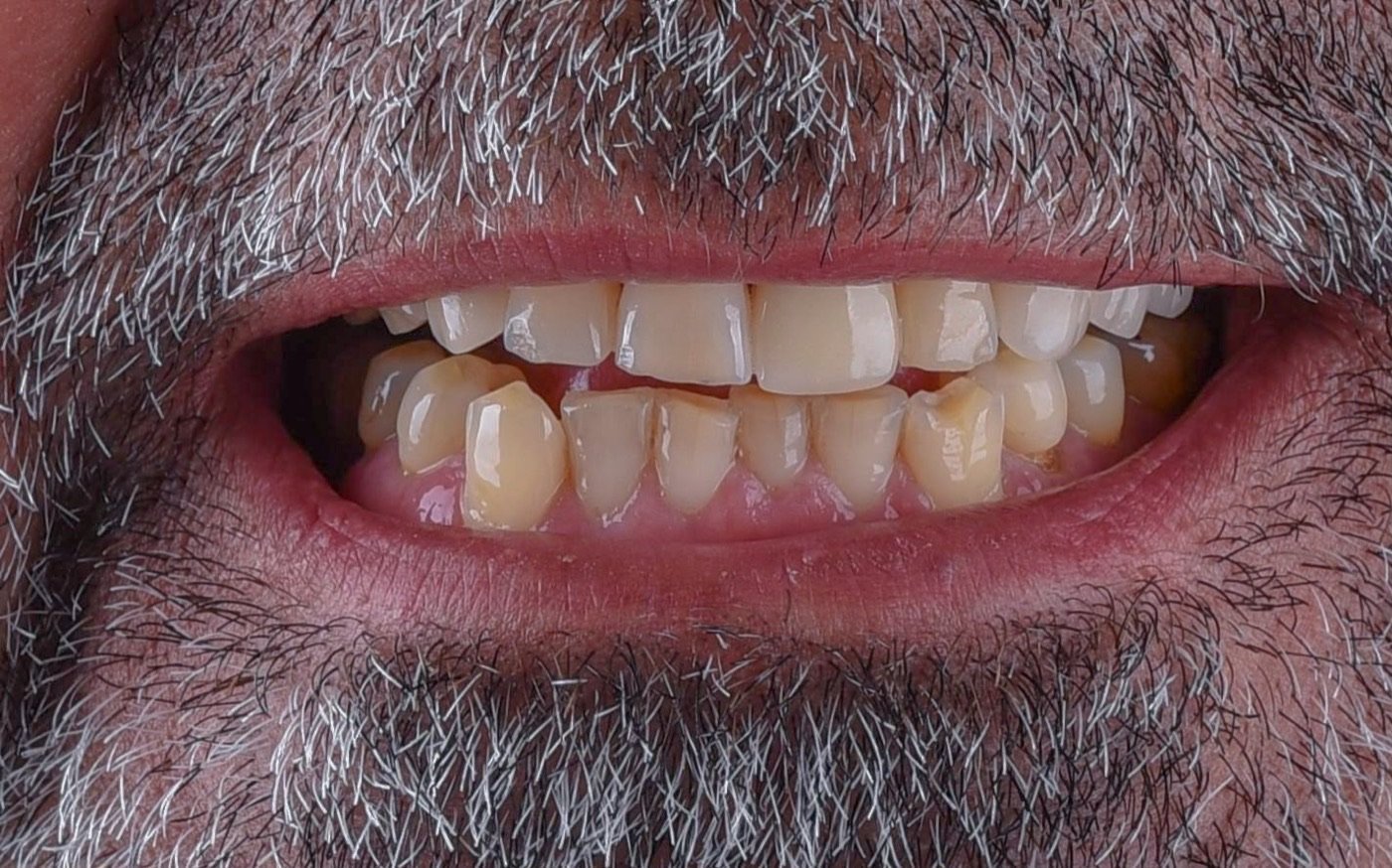
The upper front teeth had yellowed and their edges were chipped and uneven, so the smile line stepped up and down rather than following the lower lip. The lower front teeth were crowded and overlapping, and their gum levels sat at visibly different heights from one tooth to the next.
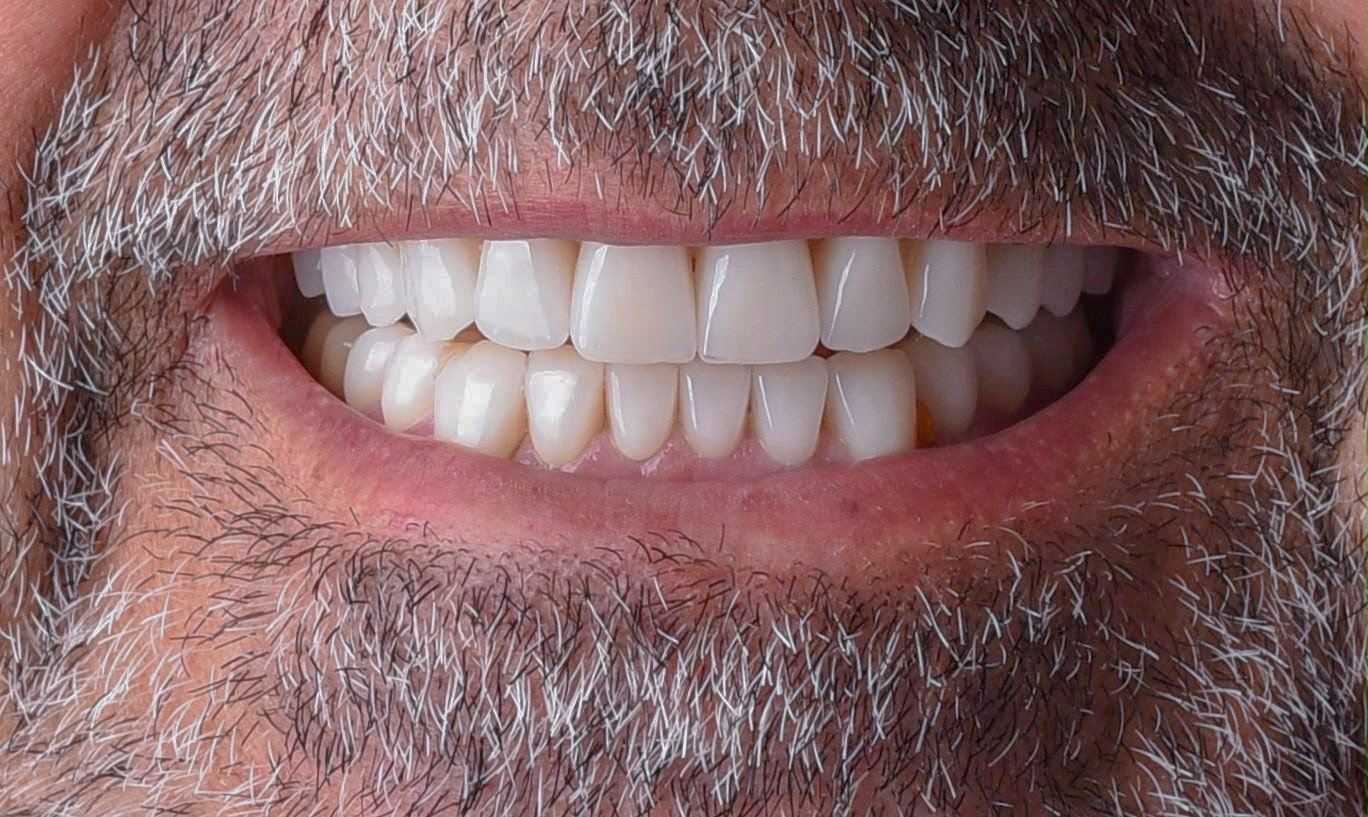
That last point drove the sequence. The gum line at the lower front teeth was levelled before any ceramic was fitted, because ceramic finished against an uneven gum margin locks that unevenness in permanently — and no amount of good laboratory work hides it afterwards. It is the sort of detail nobody consciously notices in a smile, and the reason a result can look subtly untidy even when every tooth has been corrected.
Two materials were used, chosen by position. The front teeth are lithium disilicate, which passes light the way enamel does and is what makes a front tooth read as a tooth rather than a chip of white. The back teeth are zirconia, which is considerably stronger under chewing load, and where optical subtlety matters much less than durability. One material throughout would have meant accepting a compromise at one end of the mouth or the other.
Both photographs are natural smiles taken from the same angle, so this is a like-for-like comparison.
 Before
 After
After
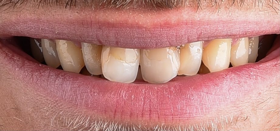
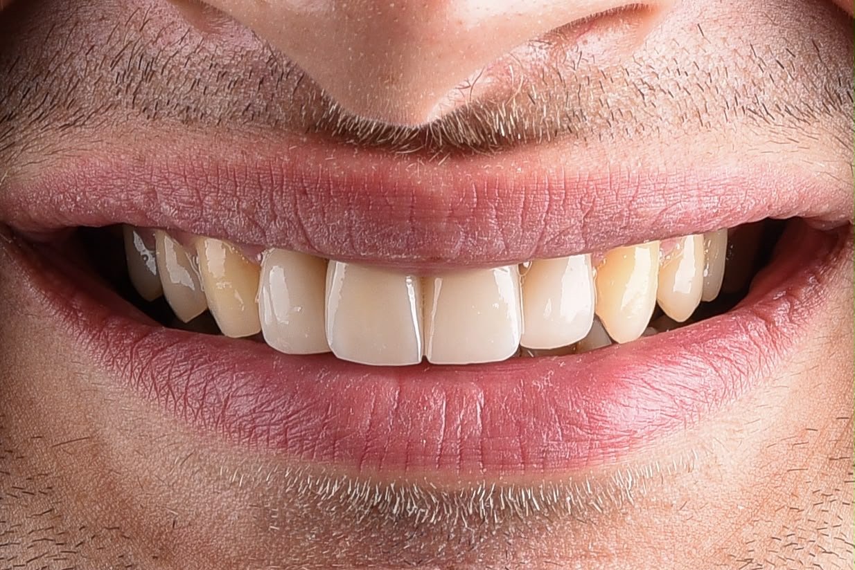
This was, honestly, a borderline case for veneers, and it is worth saying so. It reached us as a referral from an orthodontist, and the patient is young — which changes the calculation entirely. Enamel does not grow back. Someone this age will carry whatever is done here for decades and will need it renewed at least once in that time, so every fraction of a millimetre left in place now is an option kept open later.
Before treatment, one of the upper central incisors was an older restoration in an opaque shade that matched nothing around it and had stained along the gum line. The teeth beside it had yellowed unevenly, and the edges no longer followed a single line.
That made the brief a narrow one: correct four front teeth with the least preparation the case would tolerate, and match the result to the patient’s own teeth rather than to an idea of white. The restorations are lithium disilicate, which keeps its strength in thin sections and passes light the way enamel does — it is the material that makes minimal preparation viable at all. We took that as far as it goes.
Which is why the canines and the teeth behind them still carry their natural, warmer colour, and why you can see a step in shade where the work ends. That step is the intended result, not a compromise: making this mouth read as one uniform white would have meant cutting far more tooth than a patient this age should have cut. Both photographs are natural smiles.
 Before
 After
After
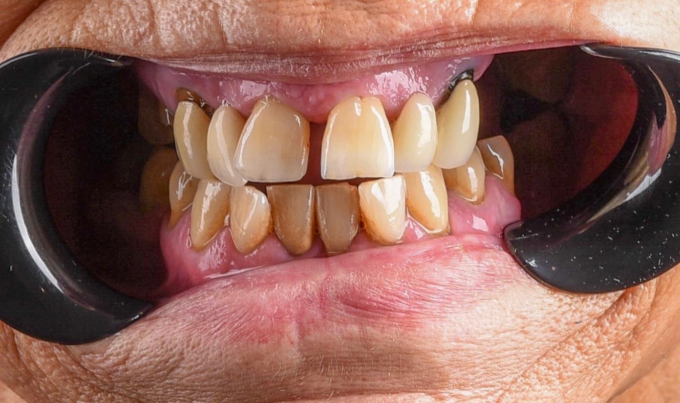
This patient is older and had been missing back teeth for years — more common than it sounds, and more consequential than it looks. When the back teeth go, the front teeth start doing a job they were never shaped for. They carry the whole load of chewing, they wear down, and as they shorten the distance between the jaws closes with them. The first photograph is where that ends: upper and lower front teeth meeting almost edge to edge, worn flat and shortened unevenly, and empty space behind them on both sides.
The temptation with a mouth like this is to treat what shows. Crowns on the front teeth would have photographed well and solved nothing, because the reason those teeth were worn out is that nothing behind them was sharing the work. New ceramic on the front of a collapsed bite wears out too — faster, and more expensively.
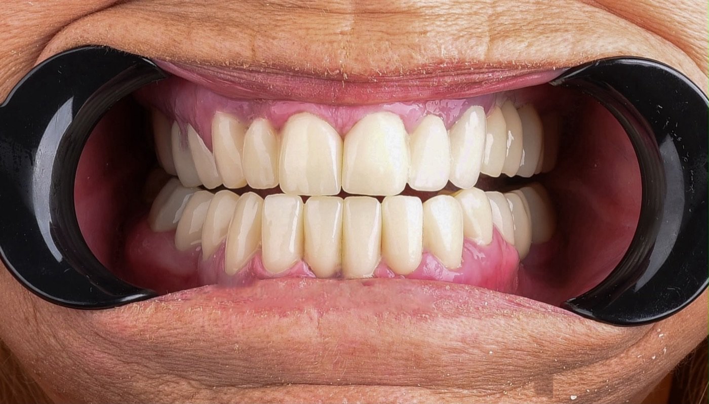
So the sequence ran the other way round. The back teeth were restored first, which is what re-establishes contact between the arches and gives the jaw relationship back to the muscles and joints that have to live with it. Only once the bite was being carried at the back were the front teeth rebuilt, to a length that belongs to the restored jaw relationship rather than the collapsed one.
The result is easier to eat with than it is to photograph, and that is the right way round. Appearance follows a mouth that works; it does not substitute for one. At this patient’s age the difference between a bite that functions and one that merely looks tidy is the difference between eating properly for the next fifteen years and not. Both photographs are retracted views taken the same way, so the change is directly comparable.
 Before
 After
After
Teeth slightly apart
 Before
 After
After
Natural smile
 Before
 After
After
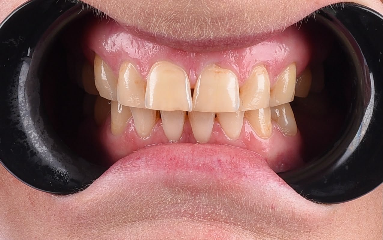
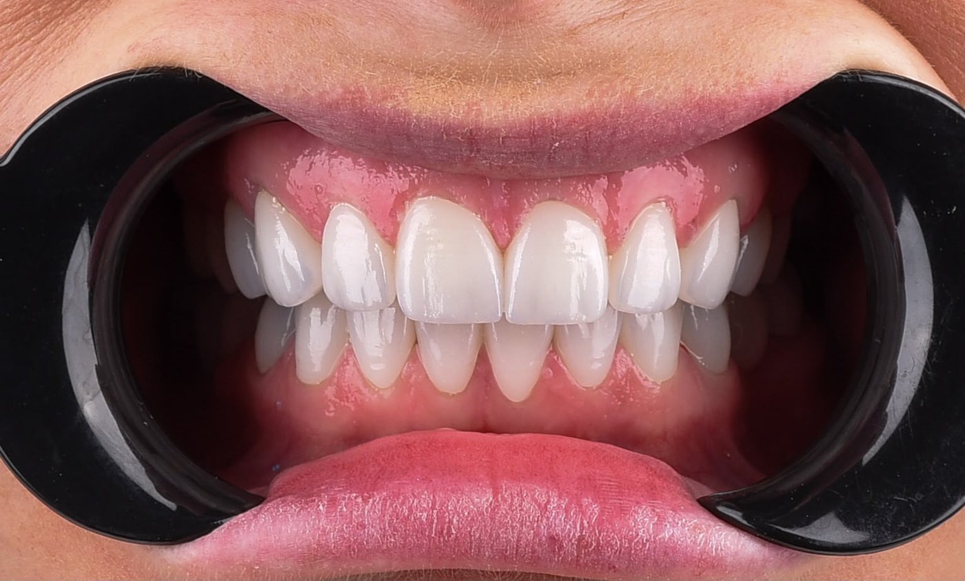
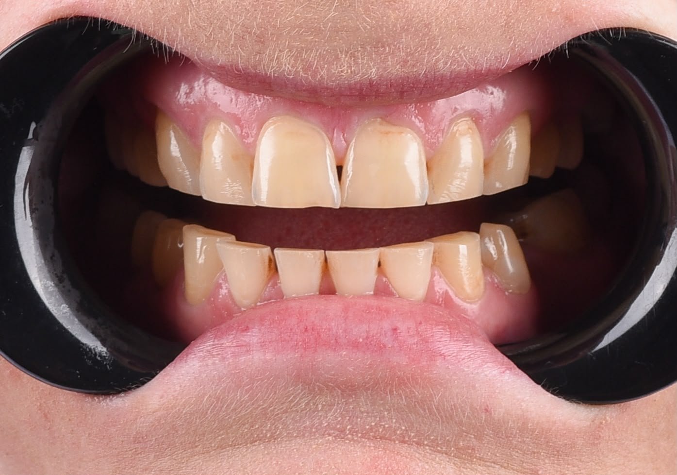
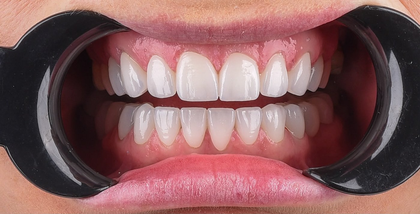
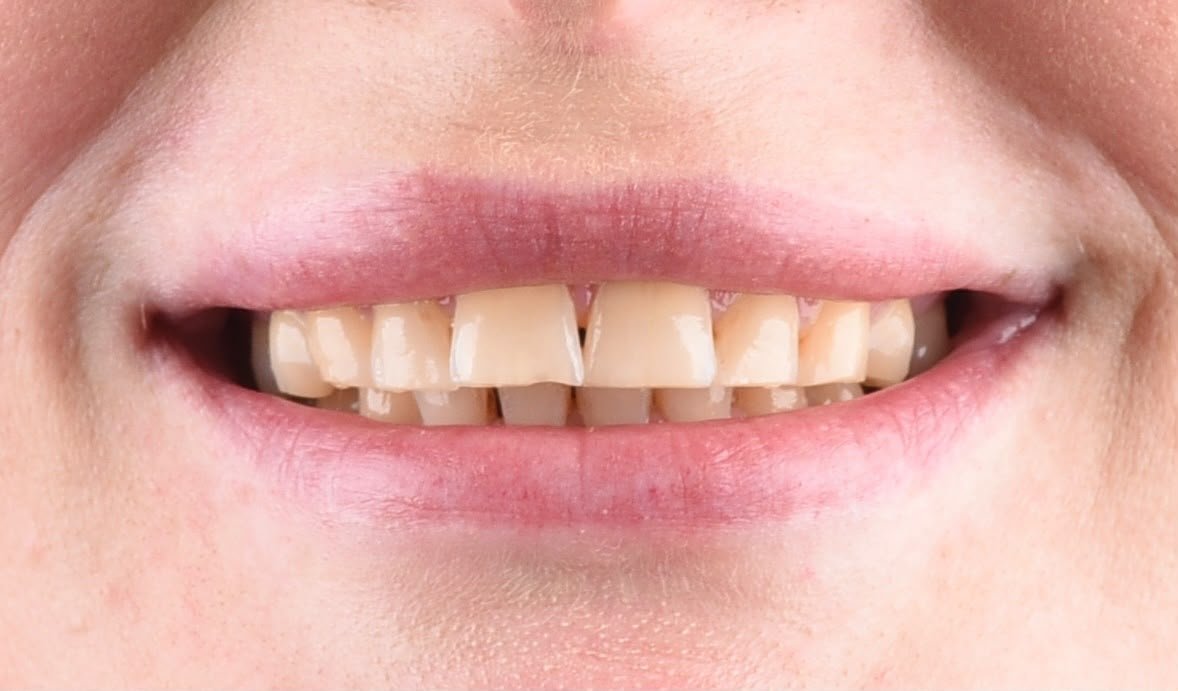
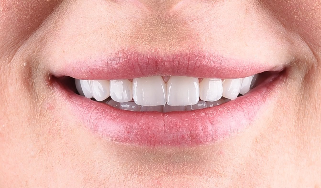
Three things were wrong at once, and fixing any one of them on its own would have left the other two looking worse. The teeth had darkened to a deep yellow across both arches, with brown staining collecting along the gum line and in the spaces between them. The upper central incisors had a small gap between them, and the lower front teeth had small gaps of their own. And the edges were worn: the upper front teeth no longer finished on one line, and the lower ones had been ground flat.
Whitening would have lightened the colour and left the gaps and the worn edges exactly where they were. Staining this deep, much of it at the gum line, also rarely lightens evenly. Closing the gaps with bonding alone would have meant adding material to teeth that were already the wrong length and shape. So the work was planned as one restoration of both arches. Shape, length, spacing and colour were decided together, across every tooth that shows when this patient talks and smiles, upper and lower.
The result is a light shade, and that deserves a sentence of its own. How light is something we decide with the patient before anything is bonded, and it is the one decision in a case like this that cannot be cheaply reversed afterwards.
This case was photographed three ways: teeth together with cheek retractors, teeth slightly apart, and a natural smile. Each before and after was taken the same way, so every pair can be compared directly.
Photographed at the clinic and published with each patient’s written consent. Every mouth is different: what is achievable for you depends on your bone, gums, bite and starting shade, and can only be established after an examination. These images show what these patients achieved — they are not a promise of your result.
Most of the cases above are some combination of these three. Each page sets out what is involved, how many days it takes and what it does not fix.
Titanium roots that restore a single tooth or a whole jaw, placed with 3D-guided planning.
A complete fixed arch of teeth supported by four or six implants — often fitted within days.
Wafer-thin ceramic shells bonded to the front of your teeth to reshape colour, form and alignment.
A dentist here will look at it and tell you what is realistic — what can be treated, roughly how many days it needs and what we would not recommend. No charge, no obligation.